By Darren Tran and Peter R. Mouton
—
Incorporating stereology into your research can be major investment, especially when purchasing a Complete (Turnkey) Stereologer system. A cost-effective alternative is equipping an existing microscope with the necessary objectives and hardware for computerized stereology. Here we outline the various components for computer-assisted microscopic analysis of biological tissue using unbiased methods.
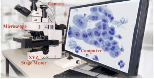
Figure 1: Hardware requirements for all computerized stereology systems.
As shown in Figure 1, all computerized stereology systems require four hardware components:
1. Microscope.
Whether brightfield or fluorescence, upright or inverted, a microscope serves as a stand for holding objective lenses to magnify images at low, middle and high resolutions. Reference spaces, i.e., anatomically defined regions of interest, are outlined at low magnification (2x, 4x), whereas high magnification stereology analysis is done with thin focal planes using ~60x and 100x lenses for cellular and subcellular structures, respectively.
2. X-Y stepping motors and Z-axis focus drive and linear encoder.
Stepping motors either built into the microscope or added as an external attachment, allow for unbiased sampling of stained tissue sections in the XY plane. The combination of z-axis focus motor and linear encoder guarantees accurate measurement of section thickness and unbiased sampling through a known z-axis distance in your tissue sections.
3. Digital camera system.
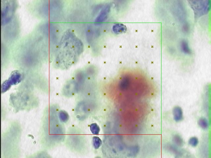
A wide variety of cost-effective cameras allow users to visualize and capture digital images for manual and automatic collection of stereology data, such as amyloid load from transgenic mouse models of Alzheimer’s disease (Figure 2).
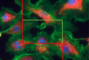
Figure 2: Computerized stereology permits users to sample and analyze biological structures in an unbiased manner (Mouton et al., 2009).
4. PC running current operating system (Windows 10).
Running the stereology software on computers with above average CPU speed (Intel Core i7 processor or higher) and increased memory (16GB or higher) will allow for faster data collection. Consequently, this will lead to large numbers of stored images which will require HD storage with a minimum of 500 GB and solid state drives (SSD).
Cited Work and Additional Reading:
P.R. Mouton. Principles and Practices of Unbiased Stereology: An Introduction to Unbiased Stereology. Johns Hopkins University Press; 1st Edition (May 16, 2002).
P.R. Mouton, M.E. Chachich, C. Quigley, E. Spangler, D.K. Ingram. Caloric Restriction Attenuates Cortical Amyloidosis In A Double Transgenic Mouse Model Of Alzheimer’s Disease. Neuroscience Lett 464:184-7, 2009.
P.R. Mouton. A Concise Guide to Unbiased Stereology,” Johns Hopkins University Press; Illustrated Edition, August 15, 2011.
M.J. West. Basic Stereology for Biologists and Neuroscientists. Cold Spring Harbor Laboratory Press; 1st Edition, August 31, 2012.
P.R. Mouton. Neurostereology: Unbiased Stereology Of Neural Systems. Wiley-Blackwell Press; 1st Edition, February 3, 2014.
S. Alahmari, D. Goldgof, L. Hall, H.A. Phoulady, R.H. Patel, P.R. Mouton. Automated Counts of Stained Cells by Deep Learning and Unbiased Stereology. J Chem Neuroanatomy, 96:94-101, 2019.


