Intuitive and Efficient Analysis of Tissue using Unbiased Methods
Background: Manual counting in stereology is considered the gold standard within quantitative histopathology allowing histotechnologists to manually page up/down through a z-stack marking objects of interest to which the degree of movement is determined by the section thickness specified in the study parameters. [1] However, in the recent years digital pathology with the use of image analysis software is emerging for quantifying pathological biomarkers.
Through efforts to make manual counting more efficient as well as allow for a record of the z-stack analysis to be taken, SRC Biosciences has developed the Digital Disector Tool, the first approach that allows for time modulation of the stereology study to be set for efficiency while maintaining unbiased stereology principles be applied.
This expansion of manual counting to a digital (video) format will allow remote tissue analysis without a stereology system needed during analysis, time modulation of the study to be performed at a custom frames per second, and a record of study parameters quantified: cells counted, coordinates of the cells, image dimensions.
How does the Digital Disector work?
The Digital Disector is user friendly annotation tool to accelerate data collection process by speeding up stacks’ images display automatically. Thus, users will be able to access the software locally and perform tissue analysis remotely from their labs.
Acquiring Case Images (Stereologer Software)
First, the user must use the Stereologer software, stereology software sold by SRC Biosciences, to acquire case images of each z-stack. Stack parameters such as steps size, number of sections, disector height and magnification parameters for the region and object must be set. The user will then prompt the program to collect case images.

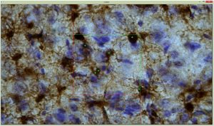
Figure 1: User-view during annotation of objects on interest.
Tissue Analysis (Digital Disector Tool)
Prompted by the Digital Disector, the user will select which case z-stack of images to perform analysis on. The user will also set a video speed (frames per second). After the user starts the analysis, a video of the images collected is played where the user can mark by clicking with their cursor the locations of the object of interest. The video speed can be increased/decreased or paused to the user’s need.
How are the results of the Digital Disector Used?
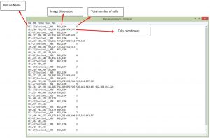
After finishing the study, the end-user will receive a text file with the mouse name, image dimensions, total number of cells, and cell coordinates. The end-user can use this information to determine the pervasiveness of the pathological biomarker through out the tissue in their histological study which have applications to neuroscience, cardiology, and respiratory studies. Data can also be stored for reporting, collaborative review, etc.
Figure 2: User-view of study results
For more information, see our stereology software page: https://srcbiosciences.com/stereologer-software
Reference: Mouton, P.R. (2002) Principles and Practices of Unbiased Stereology: An Introduction For Bioscientists. The Johns Hopkins University Press, Baltimore, MD.


