Stereology Systems
Incorporating Unbiased Stereology into Your Research
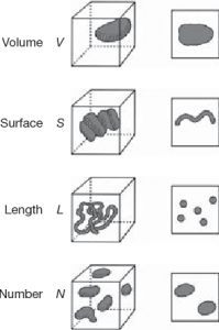
Stereology uses unbiased sampling and estimation methods to quantify tissue with first-order stereological parameters such as – volume (V), surface area (S), length (L), and number (N) [1] as seen in Figure 1. This allows for quantification of structures in an unbiased (aka design-based), rather than assumption- or model-based approaches.
Modern stereology is typically applied to studies involving stained cells where structural changes such as cell loss, synaptic degeneration, loss of volume (atrophy) is quantified [2]. Eliminating or avoiding all known sources of bias leads to theoretically unbiased estimates, i.e., results that converge on their expected or true values as tissue sampling continues. When bias is present, the results converge on values some distance from the true value.
This application is made through stereology systems, which are made up of stereology software interfaced with peripheral microscope hardware.

Applications for Stereology Systems
Typical users of computerized stereology system such as Stereologer® include students and bioscientists in academia centers, government labs and pharmaceutical companies. A few examples of these applications include:
Neuroscience
Common endpoints are number and size distribution of glial cells, neurons, axons, and synapses. Studies of these structural parameters help to understand the neurobiological basis for normal functions and behaviors (e.g., learning and memory) from early development to late life. Studies of neurological diseases can include quantification of neuropathology such as neurofibrillary tangles, senile plaques (Aß load), neuroinflammation along with neuron and synaptic degeneration.
Drug Discovery
Stereology is commonly used to assess the safety and effectiveness of treatments for diseases. An example might be quantification of cell and fiber degeneration in the nigrostriatal pathway to access novel strategies for the therapeutic management of patients afflicted with Parkinson’s disease.
Cardiovascular Studies
Quantify numbers and sizes of capillaries, cardiomyocytes, and cardiomyocyte nuclei for testing hypotheses such as: “Is there a significant loss of cardiomyocytes during ventricular hypertrophy to heart failure?” or “Does a specific treatment reduce the degree of fibrosis in the heart?”
Pulmonary
Typical stereology studies focus on quantifying alveoli number and surface are in the lungs to access trauma, toxicity, disease, and genetic aberrations. Other applications include volume of alveolar septal tissue, surface of the alveolar epithelium and the length of nerve fibers innervating the conducting airways.
Adding Stereologer to your research is a one-time purchase. There are no annual usage fees, and you receive continuing support & maintenance our experts at no cost. For more information reach out to us directly (Contact Us) or register online for the next class in our no-cost monthly Stereology School.
What Stereology Systems Do
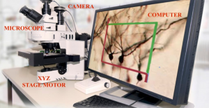
Stereologer is an integrated workstation for collecting stereology data from stored images (digital stereology) or directly from stained tissue sections (real-time stereology). The Turnkey Stereologer system includes the most current version of the Stereologer software interfaced with peripheral hardware. SRC Biosciences also specializes in building stereology systems using your laboratory equipment, e.g., microscope with motorized XYZ stage, camera, PC-based computer/monitor, as detailed below.
Components of a Stereology System
Most of the research and clinical labs have the hardware for computerized stereology, including a microscope with low to high optics equipped with X-Y stepping motors with a z-axis focus drive; a digital camera, and a PC running a current operating system.
The microscope serves as an integrated producer of image for the computer to project onto a monitor for analysis by a trained technician. Users outline regions of interest (reference spaces) and set magnifications according to the anatomical boundaries of the biological structures of interest.
The X-Y stepping motors and Z-axis focus drive automatically perform unbiased sampling of tissue sections through regions of tissue (reference spaces).

Stereology System Hardware
Microscope
Microscopes in stereology systems allow for stage movement in the XYZ positions, tracking of cell location, and speed modulation during analysis. Stereologer is compatible with all microscopes, for example, Zeiss, Leica, Olympus, and Nikon. Many modern microscopes include an internally controlled motorized XYZ stage control. If a microscope is missing this required hardware, we add them as external devices from reliable companies such as Applied Scientific Instrumentation, Prior Scientific and Ludl.
Camera
During the potential client’s initial inquiry, hardware specialists determine if a client’s current camera system is compatible with the stereology software. Companies that specialize in stereology systems maintain relationships with many camera vendors such as Zeiss, Nikon, Olympus, and Leica to determine the best camera for a lab’s research goals and budgetary limits. All third-party cameras follow industry standards such as DCAM and USB3.
Computer
All new PC computers support the Widows 10 OS and include PCIe boards for plug-in support of cameras and motorized stage connections. Preferred configurations for Stereologer systems include Intel Core i7 processor and memory of 16 GB or higher. As the needs for processing images increase with automatic stereology approaches (see below), preferred HD storage is minimum 500 GB with Solid State Drives (SSD).
Stereology System Software
General stereology software requirements include the ability to set study parameters, acquire tissue images, perform data collection, and output study results. General parameters to be set are XYZ step size, number of sections, section sampling interval, etc. Images will be captured within a set tissue thickness. These images will be analyzed using stereology probes, each company will have their own probe designs.
The Stereologer automatically determines optimal study parameters, acquires tissue images, guides data collection and outputs study results to saved data files. Below are methods available to users using SRC Bioscience’s stereology software.
Conventional Stereology
Manual counts on real-time images, rather than on digital images. In the mid 1990s, the Stereology Resource Center, Inc (now SRC Biosciences, Tampa, FL), in collaboration with the Johns Hopkins University (Baltimore MD) and Applied Physics Lab (Laurel, MD), developed and commercialized the Stereologer®, the first fully integrated, comprehensive computerized system for unbiased stereology. This system continues to lead the industry through state-of-the-art innovations for more accurate, user-friendly, and increasingly automatic collection of stereology data from tissue sections and images.
Digital Stereology™
Digital Stereology™ refers to the automatic unbiased sampling and manual analysis of z-axis stacks of digital images, i.e., disector stacks. Digital images provide the data for analyses with the ability to archive permanent evidence of collected data.
There are two options for stereological analysis of digital images:
- “Slice-wise” manual counts. The user scrolls through each z-stack in real-time using the up/down arrows while counting (clicking) on the stained objects of interest.
- Digital Videos. Each 3D stack of z-axis images is automatically converted into a digital video and presented for rapid data collection at speeds controlled by the user.
Automatic Stereology (Fully Automatic Stereology Technology, FAST®)
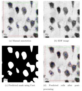
Deep learning, a form of artificial intelligence (AI), is being incorporated into all disciplines of scientific research including the Stereologer system.
SRC Biosciences received grant funding from the National Science Foundation to develop the first AI-based approaches to accelerate the accurate and reproducible collection of data using unbiased methods [3].

The Best Stereology System?
Stereologer is widely considered the best stereology system in industry and academia from several points of view.
- Stereologer is the only computerized stereology system in the industry that allows manual, semiautomatic, and fully automatic analysis of stained tissue sections.
- The system software includes an intuitive design that allows for efficient and accurate analysis of stained tissue sections and stored images from brightfield, fluorescence, confocal, and electron microscopes.
- The user-friendly Stereologer software interfaces with the widest range of hardware components.
- The Stereologer system is far less expensive and delivers higher performance than more costly systems.
- Due to high design standards developed over the past three decades, we can sell and support Stereologer for the lowest price in the industry and you pay no further costs for as long as you own the system. In other words, you buy Stereologer and say goodbye to annual usage fees, access charges and high support costs charged by other companies.
Contact SRC Biosciences
For future Stereologer users, the critical question is whether you prefer a Complete Turnkey Stereologer System; or to integrate the latest version of the Stereologer software with your current hardware. The requirements for purchase of your Stereologer system will be assesses by our support team to develop a quote that fits the needs of your research program, without wasting time resources on unnecessary components or features. Contact SRC Biosciences online to learn more.
References Cited
- Mouton, P. R. (2002). Principles and Practices of Unbiased Stereology: An Introduction for Bioscientists (1st ed.). Johns Hopkins University Press.
- Brown D. L. (2017). Bias in image analysis and its solution: unbiased stereology. Journal of toxicologic pathology, 30(3), 183–191. https://doi.org/10.1293/tox.2017-0013.
- Alahmari, S., Goldgof, D., Hall, D. Phoulady, H.A. Patel, R., Mouton, P.R. Automated Counts of Stained Cells by Deep Learning and Unbiased Stereology. J Chem Neuroanat, 96:94-101, 2019.
