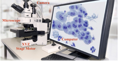
Turnkey Stereologer® System
A Complete or “Turnkey” Stereologer system includes all hardware and software for stereological analysis of stained tissue sections and stored images from brightfield, fluorescence, confocal, and electron microscopy, including large virtual slice images.
This Stereologer system includes the following components in a single quote (well within the budget of most single investigators):
Hardware Components for a Turnkey Stereologer® System include:
- Your preference of upright or inverted microscope in partnership with industry-leading microscope providers.
- Camera system to deliver image analysis capabilities tailored to your needs.
- Slide micrometer for lens calibration.
- Installation services to verify stage/focus communication, live camera display, lens calibration and preliminary systems overview.
For more information on compatible hardware with our Stereologer® system, contact SRC Biosciences.
Stereologer software for user-friendly, comprehensive, and efficient analysis of biological structures using state-of-the-art unbiased methods.

- Designed for single or multiuser basic laboratory environments focused on research in the neurosciences and other bioscience disciplines.
- Includes multi-channel analysis of multiple colocalized objects under fluorescence (UV) microscopy; automated Z-stack image acquisition for offline analysis; automatic export of data and results; and discounts on additional software licenses.
- Stereologer software training services tailored to your needs, i.e., remote via virtual platforms or on-site at your facility.
- All owners receive complimentary maintenance and support of their Stereologer software. This program (unique to the industry) provides state-of-the-art software support, without burdening owners with yearly maintenance and usage costs.
Features and functionality developed specifically for the Stereologer software:
- Current version of Stereologer software installed on a new computer for high-resolution computerized stereology, fully integrated with microscope and camera.
- Space Balls™ for unbiased analysis of length/length density of curvilinear object, e.g., fibers, axons, dendrites, capillaries, etc. (Mouton et al., 2002).
- Rare Event Protocol™ to standardize data collection of rare events, i.e., objects with extremely limited distribution (Mouton 2011).
- Virtual Cycloids™ for unbiased analysis of surface area and length, respectively, on arbitrary sections (Gokhale et al., 2004).
- Automatic workflow optimization to collect data with maximum efficiency (Alahmari et al., 2019).
- Digital Stereology™ for automatic capture and manual, semi-automatic and fully automatic stereology analysis of tissue using unbiased methods (Mouton et al., 2017)
- Integrated Training Module to ensure consistent data collection across multiple investigators.
- Integration with FIJI/ImageJ software to automatically stitch 2D captured images and create super-high-resolution images and automatically build 3D images stacks.
- Consultation services regarding preferred histology and tissue preparation methods for accurate stereology.
- Detailed User Guide covering not only how to use software but also relevant stereology principles (Mouton 2002, 2011).
For more information on Stereologer software and our upcoming upgrades for manual and automatic approaches in development, contact SRC Biosciences.

Cited Works
Mouton, P.R., Gokhale, A.M., Ward, N.L., West, M.J. Stereological Length Estimation Using Spherical Probes. J Microsc 206: 54-64, 2002.
Mouton P.R. Principles And Practices Of Unbiased Stereology: An Introduction For Bioscientists. The Johns Hopkins University Press, Baltimore MD, May 2002.
Gokhale, A.M., Evans, R.E., Mackes, J.L., Mouton, P.R. Stereological Estimation Of Surface Area In Thick Transparent Sections Of Arbitrary Orientation Using Virtual Cycloids. J Microsc 216(Pt1): 25-31, 2004.
Mouton, P.R. Unbiased Stereology: A Concise Guide. The Johns Hopkins University Press, Baltimore MD, August 2011.
Mouton P.R., Phoulady HA, Goldgof D, Hall LO, Gordon M, Morgan D. Unbiased Estimation Of Cell Number Using The Automatic Optical Fractionator. J Chem Neuroanat. 80:1-8, 2017.
Alahmari, S., Goldgof, D., Hall, D. Phoulady, H.A. Patel, R., Mouton, P.R. Automated Counts of Stained Cells by Deep Learning and Unbiased Stereology. J Chem Neuroanat, 96:94-101, 2019.
